


A wonderful band-aid.
#echography
#science
#invention
#blogpost
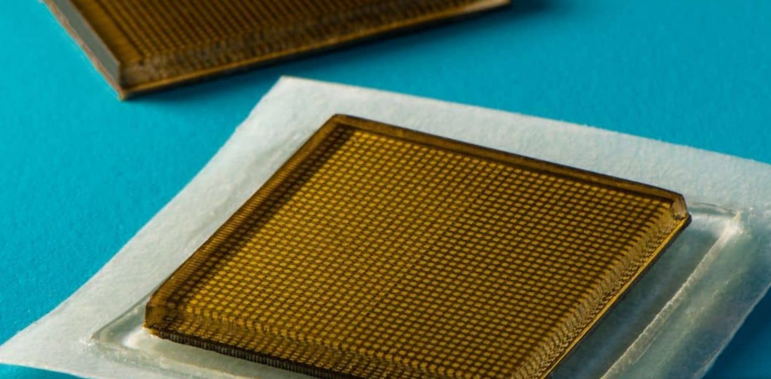
Medical researcher Xuanhe Zhao almost jumped a hole in the air when, together with his design team, he finally managed to create an effective ultrasound device. It is no larger than a stamp and it sticks sufficiently to the human.
Once on it, you could just see through the skin of a human being.
Wherever you put it, it immediately shows an image of the organ that is present underneath it.
“A new era of wearable imaging is now unlocked,” says a colleague. From the inner pleasure he has about it, his face turns a little rosette and his eyes shine like green apal stones.
Normally, before an ultrasound, you must first apply a liquid gel to a patient's skin. After that, an ultrasound head or tranducer is placed on your body.
The echo head then emits sound waves. These are reflected back by the organ in your body. These echo signals are then converted into an image.
With the old method, the gel dries out after a while. Then you won't see anything at all.
It is also difficult, even with a robotic arm, to keep the transducer in the desired position for a long time
With the miniscule patch, this problem is solved.
A lot of couples know the former device. Full of anticipation, with a big belly, they look at their cute tiny fetus on the screen.
The newly developed patch sticks to the skin thanks to two thin layers of elastomer that surrounds a middle layer of solid hydrogel. It is a largely water-based material that allows the sound waves to pass through easily. In contrast to the former gel, the latter developed is elastic and stretchy.
The elastomer ensures, as you probably suspect, that the hydrogel does not dry out.
The bottom layer sticks to the skin. While the upper one attaches to tiny transducers.
The ultrasonic small emergency is about two square centimeters in diameter and three millimeters thick. Almost the same as a stamp. In addition, it can deliver clear images of inner organs for 48 hours.
The thing also sticks like the best.
Volunteers did various activities such as sitting, standing, jogging, cycling and even lifting weights.
While the researchers observed their large blood vessels, heart, lungs and stomach.
At the moment, the device still has a nuisance. It still needs to be connected to a device with wires that translates the sound waves into images.
Work is being done to transmit these images wirelessly.
Then the patches can even go home.
“We imagine a number of patches that can be applied to different places on the body,” says Zhao, “We will make sure that they can communicate with mobile phones. In addition, certain smart algorithms are incorporated to be able to analyze the images on request.
It's a huge step forward. A device that offers many new possibilities”
“This is how it can also be used by consumers,” his colleague jumps in, “They can then buy them to look at their muscles or also their internal organs, for example. This mainly benefits from sports training”
“Not to forget the detection of tumors,” Zhao continues”
“And of course also the development of the unborn fetus in the womb,” Pnon ends.
“Yes Zhao concludes, it's a real breakthrough”
His eyes also sparkle extra.
“It is feather-light and therefore easy to move and it is a great way to call up medical imaging”
.
Sharing = earning
1,000,000 views = € 1,000
100,000 views = € 100
10,000 views = € 10
1,000 views = € 1
500 views = € 0.50
250 views = € 0.25
100 views = € 0.10
50 views = € 0.05
25 views = € 0.025
10 views = € 0.01
And much more
- Comments (43)
- Recommended
- Milestones

kelly_Pan
3 years ago
it's amazing how technology and science are advancing, the bad thing is that we are not capable of healing the planet, I wish we were more effective there too, if not everything else will be worth

Maria
3 years ago
it's amazing how technology and science are advancing, the bad thing is that we are not capable of healing the planet, I wish we were more effective there too, if not everything else will be worth

Alberto
3 years ago
are inventions that manage to revolutionize the field of medicine a lot, which could make an improvement Wonderful post toto

Freli
3 years ago
A cure for different things that could be caused by skin perspiration, giving a great invention for the public

Alfred
3 years ago
This could lead to making forms of medicines by means of these prints would be a better form for medicines

Daniel
3 years ago
My question is if you already have an idea because you don't start? This would be an excellent help

armandofuen2012
3 years ago
There are some species of patches that help administer some kind of medication to people They are still in progress

Rasher
3 years ago
Certainly science has made a lot of progress, having good ideas, as it stands, the concept makes it an incredible job.! Although more progress is still needed

Meily
3 years ago
Science, its advances and something else I like everything you share with us you teach us a lot of things thank you for that!

Sela
3 years ago
This is a wonderful idea here is where you see the size of your heart friend I think it's a great idea

leid

3 years ago
There are already some patches that give medicines, but they are not miraculous and only work for a certain disease, hopefully they will be invented.

devian
3 years ago
Oh yes this would be very good for us but very bad for other businesses I think that's why they don't invent it

Mary Ortega
3 years ago
Technology is advancing at magnificent speeds to be able to reach this moment, which would help a lot today.

willbelysy
3 years ago
How incredible is the progress from day to day. Technology is a wonderful thing, every day we get much closer to that modernized life

Kshreyash
3 years ago
Evolution is powerful. I tend to underestimate the power of evolution but acknowledging this i think i must rethink on my opinion on evolution

ALBA P.R.
3 years ago
This is evolution. New things coming and old things fading. Your post is really informative. Thanks for sharing it with us.

Adri Noor
3 years ago
it is a very extraordinary advance, it will allow many things to change and facilitate more comfortable and effective examinations and evaluations

Kinderworkshops Petra
3 years ago
They invent such new things these days. You just have to come up with it. And then this. I've been reading with interest. Handsome work.

Dewaputra
3 years ago
What they don't invent. Of course, such a patch is a lot easier and more convenient than an extensive medical examination.

Gollo
3 years ago
How good it is to see how technology advances with new alternatives, I think the invention is very good, in short, wonderful.

Omoajayi
3 years ago
That is am impressive and brilliant development, this will help in a lot of medical diagnostic break through..Great development for the world

naf1971naf
3 years ago
I think it's ideal, it's a breakthrough, you know @Toto Animo while reading your publication I was thinking about the ailments that will no longer be necessary to go through, rubbing a translator device on the place where an inflamed organ is located must be very painful, with this invention that will remain in the past
roxana_ac
3 years ago
Hi Toto, sorry, I can't send the screenshot to DM. Please, check this photo, you have to search where says "Advanced" maybe in Dutch. After you click in where says Advanced, then you can open the advanced editor


Michell
3 years ago
I like the idea I as a woman can see that it would be so easy for us in everything and it would be a big step for the human being

alexandersolano091
3 years ago
your wonderful would be a great invention imagine being able to see a child's heart and hear the heartbeat with just that a band-aid

Ari
3 years ago
Wow, I like that innovative future of history, but I'm a little afraid that those chips will end up dominating our brain.

Betania202
3 years ago
it is a great publication and that if possible the existence of an instrument would be great for human beings

Naturefreak
3 years ago
And when did this great invention take place and is it already actively used, for example, in the hospital or by consumers?

Little Ekko
3 years ago
I think it's fabulous because there are great advances that are being made with respect to what technology is

Sieglett
3 years ago
it is a great achievement for human beings to see that technology continues to advance every day and everything for our well-being

Tatti Ps
3 years ago
it's a wonderful solution to everything and it's a breakthrough for technology it really would be great to have an instrument like that on hand

Leo 1
3 years ago
A wonderful story that I enjoyed reading by the way. With some minor adjustments and improvement of some types of errors, this story may stand a chance in the new review section set up by HJ. I'd check that out if I were you. You might be able to earn a lot of YP.

Rudy Siedlecki 🌳
3 years ago
Medical imaging is rapidly advancing. In the past, Antwerp's Agfa-Gevaert was the world leader in the production of film plates for X-ray photography.

dalinroma2.0
3 years ago
it would be great to have a device on hand that helps you stay healthy and see the things that your body shouldn't have
Here are your recommended items...
Here are your milestones...